
Figure 8.
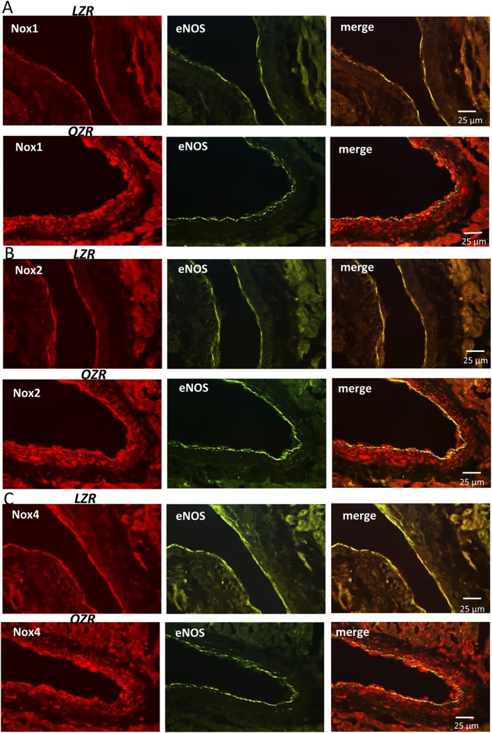
Immunohistochemical localization of Nox1, Nox2 and Nox4 in coronary arteries from LZR and OZR. Immunofluorescence for (A) Nox1 (red areas), (B) Nox2 (red areas) and (C) Nox4 (red areas) was absent or modest in coronary arteries from LZR (A,B,C left) but was markedly increased in both endothelium and VSM of coronary arteries from OZR (A,B,C, left). The endothelium was visualized with anti‐eNOS antibodies (green areas) and the double immunofluorescence shows colocalization of eNOS and Nox1 (A, right), Nox2 (B, right) and Nox4 (C, right) in the endothelium (yellow areas), but also a strong immunoreaction for all 3 Nox isoenzymes in coronary VSM (red areas) of obese rats. The sections represent n = 3 animals.