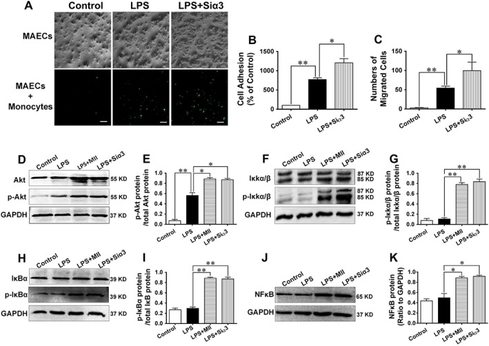
Figure 5.

Adhesion and migration of monocytes to MAECs and mechanisms underlying the effect of the α3‐nAChR antagonist α‐conotoxin MII on the inflammatory response in MAECs. (A and B) Adhesion of monocytes to MAECs after the administration of LPS (5 ng·mL−1) in the absence or presence of α‐conotoxin MII (MII, 0.1 μmol·L−1). Monocytes were incubated with an anti‐CD136 polyclonal antibody and an FITC‐conjugated secondary antibody. Pictures were taken by a using a fluorescence microscope. (C) Numbers of monocytes migrating to the lower compartment of a Transwell system. (D–K) Expressions of Akt, p‐Akt (Ser473), IκKα/β, p‐IκKα/β (Ser176), IκBα, p‐IκBα (Ser32) and NFκB were detected by performing western blotting analysis after MAECs were stimulated with LPS in the absence or presence of MII or after cells were knocked down with the gene of the α3‐nAChR. Scale bar = 100 μm. Values are means ± SEM. Data were obtained from six separate experiments (n = 6). Significance of the difference between groups is indicated as follows: * P < 0.05; ** P < 0.01.
