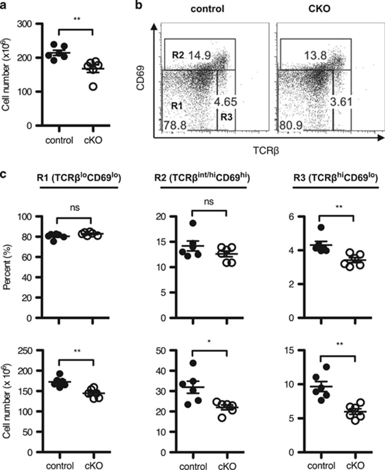
Figure 3.
TCR-experienced thymocytes are decreased in Twist2-deficient mice. (a) Thymic cellularity in control and cKO mice is shown (error bars, ±S.E.M., n=6). (b) Total thymocytes were stained with anti-CD4, anti-CD8α, anti-TCRβ, and anti-CD69 antibodies. The representative TCRβ/CD69 plots are shown. (c) The graph presents the percentage (top) and the cell number (bottom) on the following populations in cells (error bars, ±S.E.M., n=6): R1 (TCRβloCD69lo), R2 (TCRβint/hiCD69hi), and R3 (TCRβhiCD69lo) as in panel (b)

