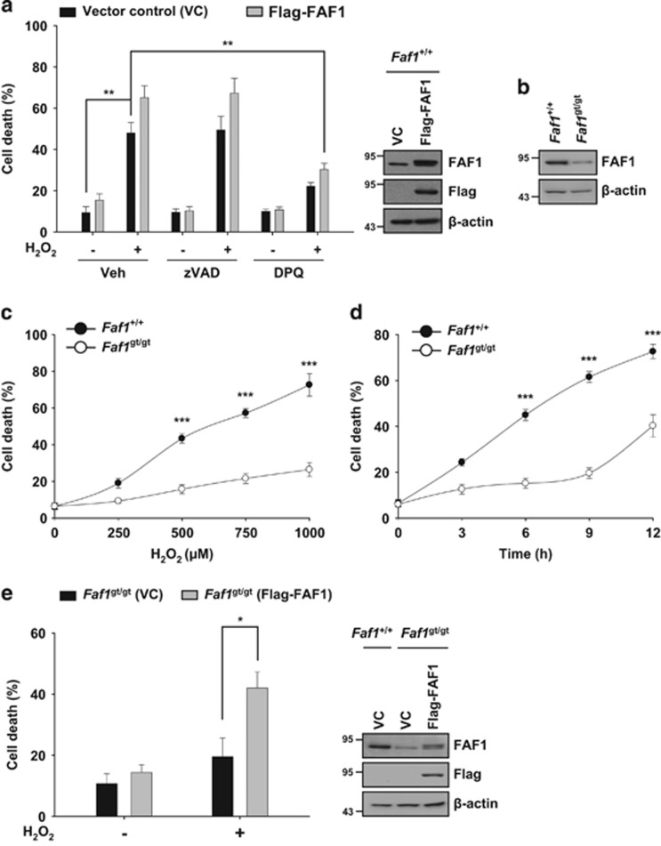
Figure 2.
FAF1 is required for PARP1-dependent necrosis after H2O2 treatment. (a) Left panel: WT MEFs were transfected with the vector control (VC) or Flag-FAF1 plasmids. At 36 h after transfection, cells were pretreated with vehicle (DMSO), zVAD-fmk (100 μM) or DPQ (30 μM) for 1 h and were then treated with 500 μM H2O2 for 6 h in presence of individual compounds. Cell death was determined on the basis of LDH release (n=3). Right panel: representative immunoblots show the levels of FAF1, Flag and β-actin expression. (b) Immunoblot analysis of FAF1 expression in immortalized Faf1+/+ and Faf1gt/gt MEFs. β-actin expression was used as an endogenous control. (c) Faf1+/+ and Faf1gt/gt MEFs were treated with the indicated concentrations of H2O2 for 6 h. Cell death was determined on the basis of LDH release (n=3). (d) Faf1+/+ and Faf1gt/gt MEFs were treated with 500 μM H2O2 for the indicated times. Cell death was determined on the basis of LDH release (n=3). (e) Left panel: Faf1gt/gt MEFs were transfected with the VC or Flag-FAF1 plasmids. Thirty-six hours after transfection, the cells were treated with 500 μM H2O2 for 6 h. Cell death was determined on the basis of LDH release (n=3). Right panel: representative immunoblots show the levels of FAF1, Flag and β-actin expression. Data (a, c–e) are expressed as the mean±S.E.M. from three independent experiments. Statistical comparisons were evaluated by ANOVA test followed by Tukey HSD (a, c–e) post hoc analysis. ***P<0.001, **P<0.01 and *P<0.05

