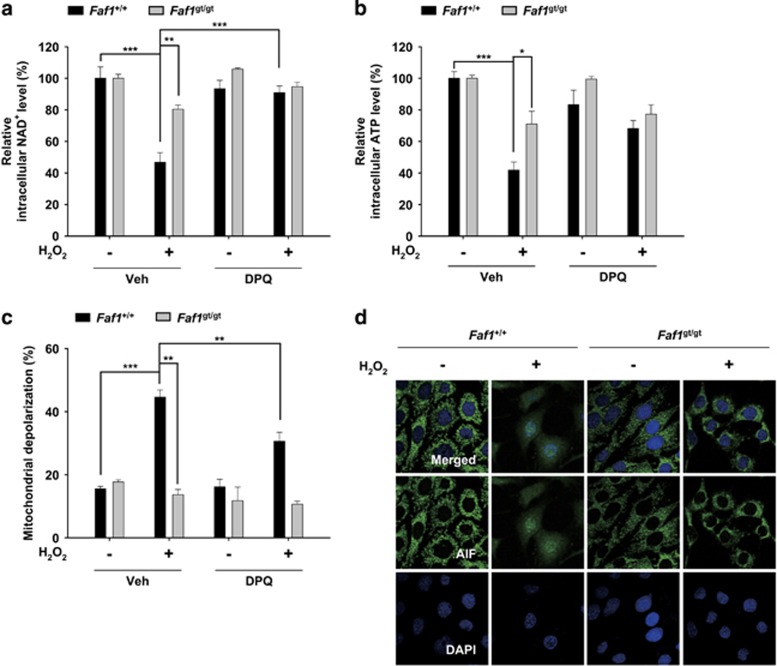
Figure 5.
FAF1-mediated PARP1 activation induces energy collapse, mitochondrial depolarization and AIF translocation during oxidative stress. (a and b) Faf1+/+ and Faf1gt/gt MEFs were pretreated with vehicle (DMSO) or DPQ (30 μM) for 1 h and then treated with 500 μM H2O2 for 1 h. Depletion of intracellular energy was determined by measuring the levels of NAD+ (a; n=3) and ATP (b; n=3). (c) Faf1+/+ and Faf1gt/gt MEFs were pretreated with vehicle (DMSO) or DPQ (30 μM) for 1 h and then treated with 500 μM H2O2 for 4 h. The cells were analyzed for mitochondrial membrane depolarization with a Muse analyzer (n=3). (d) Faf1+/+ and Faf1gt/gt MEFs were treated with 500 μM H2O2 for 4 h and subsequently immunostained with the anti-AIF antibody. The nuclei were stained using 4',6-diamidino-2-phenylindole (DAPI) and the cells were analyzed by confocal microscopy. Data (a–c) are expressed as the mean±S.E.M. from three independent experiments. Statistical comparisons were evaluated by ANOVA test followed by Tukey HSD post hoc analysis. ***P<0.001, **P<0.01 and *P<0.05

