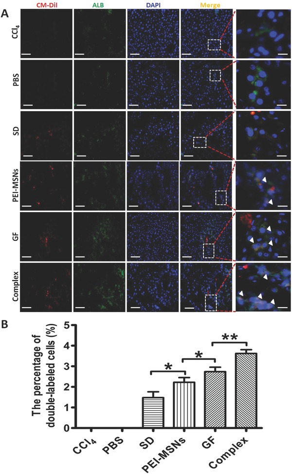
Figure 7.

Cell differentiation of transplanted mESC‐derived DE cells from the different treatments in vivo for hepatic repopulation. A) The transplanted livers were detected by fluorescence microscopy staining in the different groups. The exogenous origin of the differentiated hepatocyte‐like cells was confirmed by costaining for CM‐Dil (red) and ALB (green). The nuclei were counterstained with DAPI (blue). Scale bar: 100 μm. Inset and white arrowhead, high magnification images of differentiated cells. Representative field showing no CM‐Dil staining in a nontransplanted liver section (CCl4 or PBS). B) Quantification of differentiated CM‐Dil+/ALB+ cells shown in A. All data are represented as the mean ± SD (n = 3). *P < 0.05 and **P < 0.01.
