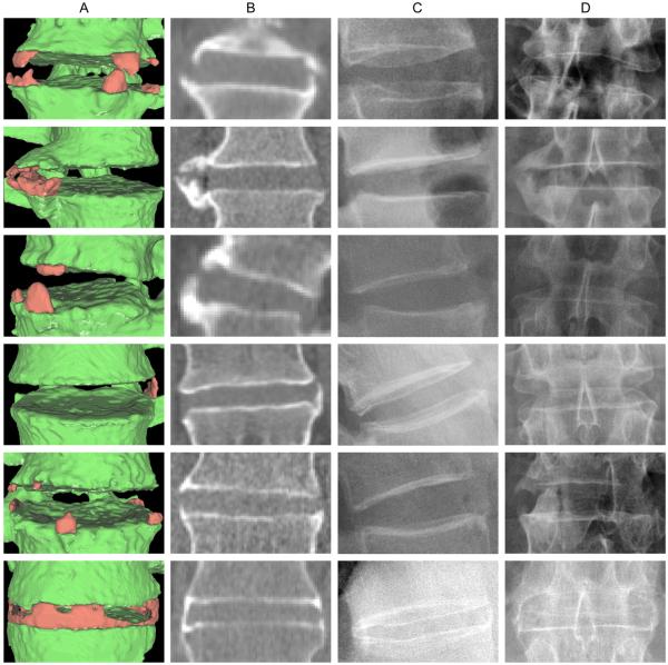
Figure 1.
Examples of syndesmophytes detected by the algorithm. Column A shows three-dimensional surface reconstructions of the CT image with syndesmophytes in red and vertebral bodies in green. Column B shows a single slice from the CT scan. Columns C and D show the corresponding lateral and anteroposterior radiographs, respectively.

