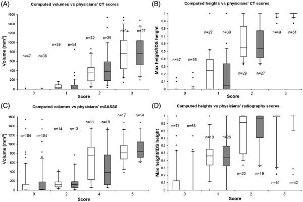
Figure 2.
Boxplots of computed syndesmophyte volume and height by physicians’ scores (white for reader 1; grey for reader 2). (A) Computed volumes versus physicians’ CT volume ratings. (B) Computed heights versus physicians’ CT height ratings. (C) Computed volumes versus modified Stoke ankylosing spondylitis spinal score (mSASSS) scores. (D) Computed heights versus physicians’ radiography height ratings. N is the number of intervertebral disc spaces. For syndesmophyte volume of the CT scans, we scored each intervertebral disk spaces (IDS) as 0=no syndesmophyte; 1=small isolated syndesmophytes involving less than a quarter of the vertebral rim and no bridging; 2=syndesmophyte involving more than a quarter of the vertebral rim or focal bridging; 3=bridging involving more than a quarter of the vertebral rim. For syndesmophyte heights, 0=no syndesmophyte; 1=tallest syndesmophyte less than half of the IDS height; 2=tallest syndesmophyte more than half of the IDS height but not bridging; 3=bridging. Lateral radiographs were scored based on the mSASSS as 0=no syndesmophyte; 2=syndesmophyte but not complete bridging; 3=bridging. We scored T11 lower to L3 upper (eight anterior corners per patient). An individual IDS could therefore be scored 0 if neither corner had a syndesmophyte, 2 if only one corner had a syndesmophyte, 4 if both corners had non-bridging syndesmophytes, or 6 if bridging was present. We scored antero-posterior and lateral radiographs for syndesmophyte height as 0=no syndesmophyte; 1=syndesmophyte less than half of IDS height; 2=syndesmophyte more than half of the IDS height but not bridging; 3=bridging.

