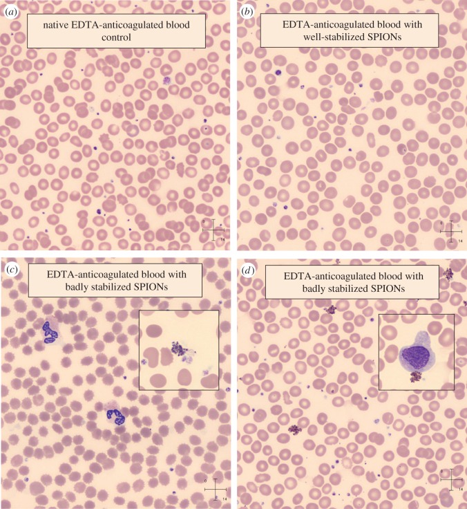
Figure 5.
Pictures of blood smears prepared from EDTA-anticoagulated blood without and with well-stabilized (a,b) and badly stabilized (c,d) SPION preparations (insets: thrombocytes, WBCs, crenated RBCs (c) and monocyte (d) and aggregated SPIONs on both). (Staining: May-Grünwald-Giemsa method by automated slide preparation system (Sysmex SP4000i); analysis: CellaVisionTM DM96. Sample description: well-stabilized (b), purified PAM-coated SPION (details in [22]); badly stabilized (c,d): (c) PEGylated oleic acid double-layer-coated SPION with high excess PEG (poly(ethylene glycol) of Mw = 1000 Da) as prepared (details in [27]) and (d) magnetic clusters of PAM-coated SPION linked by PEI (polyethylenimine) as obtained (details in [28]).) (Online version in colour.)

