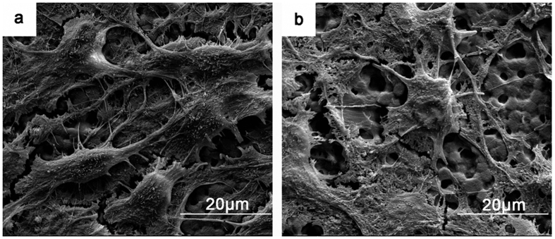
Figure 6.
SEM images of BMSC adhesion on the PPTi (a) and NPTi (b) after incubation for 7 days. The cells spreading on the PPTi were healthy and exhibited spindle and triangular shapes with numerous amount of filopodia. Less adhesion of round cells was observed on the NPTi, and the cells exhibited a collapsed morphology. These observations indicated that the PPTi was superior for cell adhesion compared with the NPTi.

