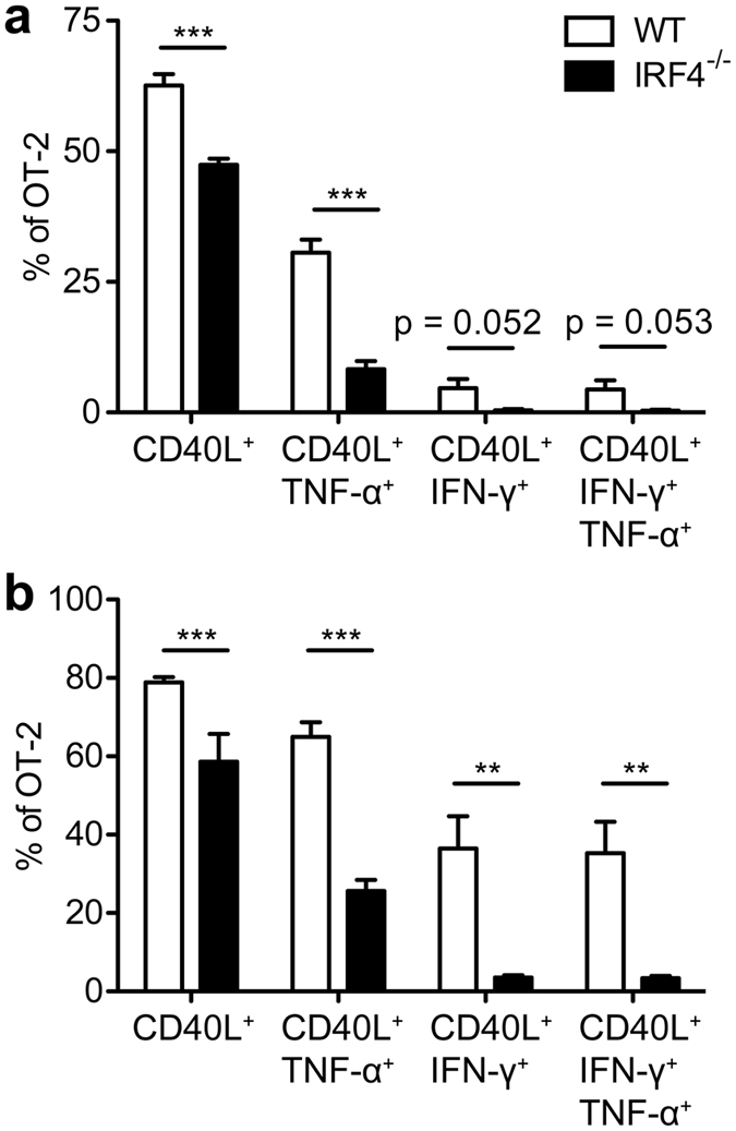
Figure 4. Reduced TH1 differentiation of transferred IRF4−/− OT-II T cells.

Purified CD4+ T cells from WT CD90.1+CD90.2+ or IRF4−/− CD90.1−CD90.2+ OT-II mice were mixed in a 1:1 ratio. 106 cells were i.v. transferred into CD90.1+CD90.2− WT mice which had been i.v. infected with 105 LmOVA one day before. On d4 post transfer, spleen cells from infected recipients were incubated for 4 h with 10−6M of OVA323-339 peptide or with PMA/ionomycin and stained intracellularly for CD40L, TNF-α, and IFN-γ. Figures show frequencies of CD40L+, IFN-γ+ and TNF-α+ cells among transferred OT-II T cells following stimulation with OVA323-339 (a) and PMA/ionomycin (b). Bars represent mean ± SEM from 6 individually analyzed mice per group. The result is representative for 4 independent experiments.
