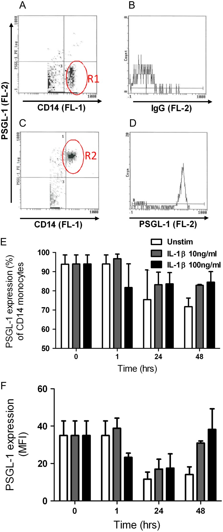
Fig. 2.
Characterization of IL-1β-induced PSGL-1 cell surface levels on human monocytes. (A) Human mononuclear cells were isolated from healthy donors (n = 2) and incubated with anti-CD14-FITC to identify the monocyte population by flow cytometry (region R1). (B) CD14+ monocytes incubated with isotype control IgG-PE to reveal background fluorescence. (C) Scatter graph revealing the percentage of resting CD14+ monocytes displaying PSGL-1 (region R2). (D) Representative histogram of CD14+ monocytes and mean fluorescence intensity of PSGL-1 levels (anti-PSGL-1-PE). hPBMCs were either treated with media alone (white bar), 10 ng/mL recombinant human IL-1β (gray bar), or 100 ng/mL IL-1β (black bar) for 0–48 hr. Cells were harvested and co-stained with PE CD162, FITC CD14 and isotype matched controls. (E) Percentage of CD14+ monocytes displaying PSGL-1. (F) Mean fluorescent intensity (MFI) of cell surface PSGL-1 levels on monocytes. Data represent mean ± SEM. This figure is available in black and white in print and in color at Glycobiology online.

