
Figure 3.
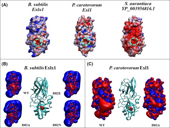
Electrostatic distribution of expansins. (A) Surface electrostatic potential of expansin structures from Bacillus subtilis, Pectobacterium carotovorum and Stigmatella aurantiaca (YP_003956816.1). The dotted circle indicates an electronegative area in the vicinity of the active Asp82 in BsExlx1 and PcExl1, which is replaced by a Leu at the equivalent position in S. aurantiaca. Colours by electrostatic potential mapped at the surface are red to blue from −5 kT/e to +5 kT/e. Isopotential contours of inactive mutants D82A and D82N expansins from BsExlx1 (B) and D82A from PcExl1 (C), showing a reduced influence compared with the electronegative Asp82 (Pastor et al., 2015).