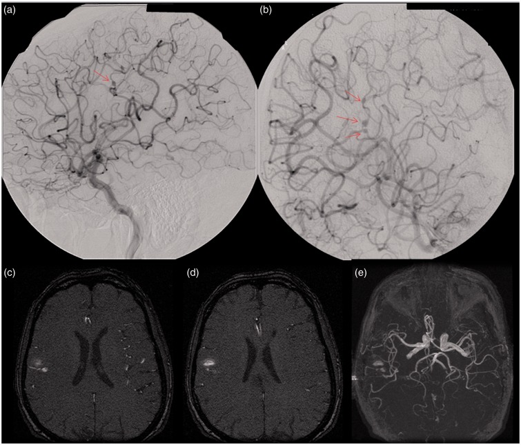
Figure 1.
This patient presented with a headache and was found to have a subarachnoid hemorrhage and native mitral valve endocarditis. Images of a right carotid injection from a right cerebral digital subtraction angiogram ((a) and (b)) demonstrate three fusiform aneurysms along the frontoparietal opercular branch of the right middle cerebral artery (arrows), best appreciated on the more zoomed-in image (b). Axial ((c) and (d)) and three-dimensional reformatted (e) images from a time-of-flight magnetic resonance angiography (MRA) demonstrate signal abnormality within the subcortical white matter of the posterior right frontal operculum secondary to hemorrhage; however, an M3 branch aneurysm is difficult to appreciate.

