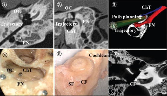
Figure 4.

The location between the drill path and surrounding anatomic structures. ① postoperative HRCT; ② the location relationship between drill path and FN; ③ postoperative verification procedures; ④ routine temporal anatomy; ⑤ the contoured cochlear; ⑥ postoperative micro-CT. The arrow shows the facial nerve. HRCT: High-resolution computed tomography; FN: Facial nerve; OC: Ossicular chain; ChT: Chorda tympani nerve; CF: Cochlear fenestration; SF: Stapes footplate; CT: Computed tomography.
