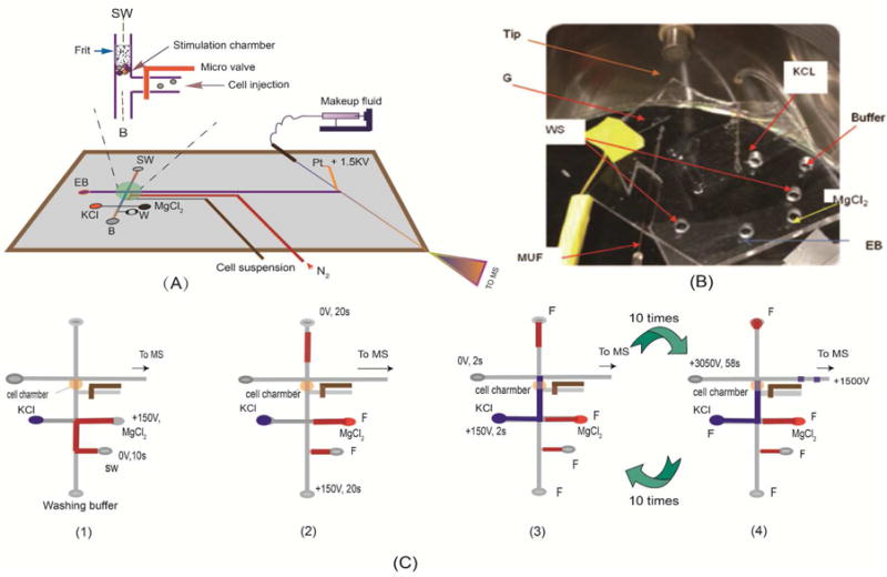
Figure 1.

(A) A schematic diagram of the proposed microfluidic platform (separation channel is 3.5cm long × 60μm wide × 25 μm deep). The nano-liter sized chamber for cell perfusion is shown in the inset; (B) a photographic image of the set-up for studying neurochemical release from neuronal cells. G is the nano-electrospray gold electrode. Other components are labeled and indicated with an arrow); (C) A schematic diagram showing the procedural operations after cells are loaded into the nano-perfusion chamber: 1) loading an inhibitor (MgCl2) solution; 2) perfusing the cells with the inhibitor solution; 3) perfusing the cells with the stimulus solution; and 4) MCE-MS analysis of the perfusate.
