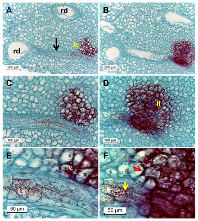
FIGURE 7.
Microscopic evaluation of resin duct contact with discolored lenticels in surface and deeper transverse histological sections of mango peel stained by safranin and fast-green after 3 weeks of cold storage. (A–F) Transverse sections of mango stored at 5°C, observed at various magnifications (A, B, magnification ×100; C, D, magnification ×200; E, F, magnification ×400) showing discolored lenticels with phenolic accumulation (stained in red) and lenticel connection to resin duct. Dense red-colored granules are seen inside the resin duct. (B,D,F) Are surface cuts and (A,C,E) are deeper cuts. rd, resin duct; lc, lenticel; black arrow, zone of mesophyll cells seen between rd and lc; yellow and red arrows, phenolic transport and accumulation, respectively.

