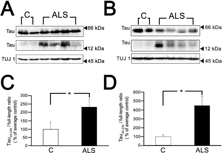
Figure 1.
High levels of tau45-230 were detected in ALS spinal cords. (A and B) Quantitative Western blot analysis of tau content in representative homogenates prepared from postmortem samples of the anterior horn of lumbar (A) and cervical (B) spinal cords from control (C) and ALS (ALS) subjects. Immunoreactive bands at 17 kDa apparent molecular weight corresponding to tau45-230 were readily detectable in ALS samples. (C and D) Graphs show the ratios of tau45-230/full-length tau in control and ALS subjects. Numbers represent the mean ± SEM of control (n = 7) and ALS (lumbar n = 22, cervical n = 10) samples. Values are expressed as percentage of controls, considering the values obtained in control subjects as 100%. Class III β tubulin (TUJ1) was used as loading control. *Differs from control subjects; P < 0.01.

