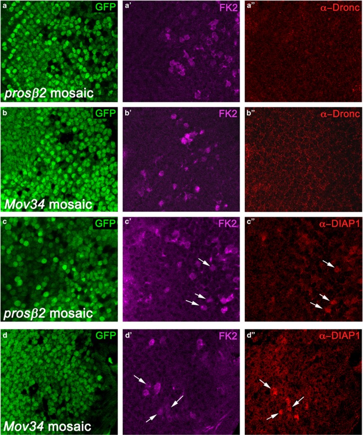
Figure 3.
Diap1, but not Dronc, accumulate in proteasome mutant clones. Shown are high magnification images (x100) of the posterior compartment of prosβ2 (a and c) and Mov34 (b and d) mosaic eye imaginal discs labeled for Dronc (a and b) and Diap1 (c and d). FK2 labeling was used to identify mutant clones. The left panels indicate the positions of the proteasome mutant cell clones by absence of GFP. In the middle panels, the proteasome mutant cell clones are positively marked by FK2 labeling (in magenta). The right panels show the Dronc (a'' and b'') and Diap1 labelings (c'' and d'') in red. White arrows mark a few cell clones as examples. See also related Supplementary Figures S3 and S4. Genotype in (a and c): ey-FLP; prosβ2EP3067 FRT80/P[ubi-GFP] FRT80. Genotype in (b and d): ey-FLP; FRT42D Mov34k08003/FRT42D P[ubi-GFP]

