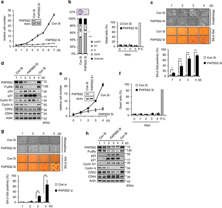
Figure 1.
PAPSS2 depletion suppresses cell growth due to premature senescence in MCF7 cells and HDFs. (a–d) MCF7 cells were transfected with Con Si or PAPSS2 Si. (a) The numbers of viable cells are shown as relative values. Total protein was extracted from cells 3 days after siRNA transfection and was subjected to WB analysis. (b) Cell cycle distributions (left graph) and dead cell populations (right graph) were analyzed by FACS 3 days after siRNA transfection and at the indicated times after siRNA transfection, respectively. Doxorubicin-treated (10 μg/ml) MCF7 cells were used as a positive control (P.C.). A colony-forming assay (CFA) was also performed (left panel) 7 days after siRNA transfection. (c) Cellular morphology and SA-β-Gal positivity were assessed at the indicated times after siRNA transfection (upper panel), and the percentage of senescent cells was quantified (lower graph). (d) Cells were harvested at the indicated times after siRNA transfection and were subjected to WB analysis. (e–h) HDFs were transfected with either Con Si or PAPSS2 Si. (e) Cell viability, (f) the percentage of dead cells, (g) morphological changes and SA-β-Gal positivity, and (h) WB analysis were performed as described in a–d. Error bars indicate the S.D. of three independent experiments. ***P<0.001, **P<0.01, and *P<0.05

