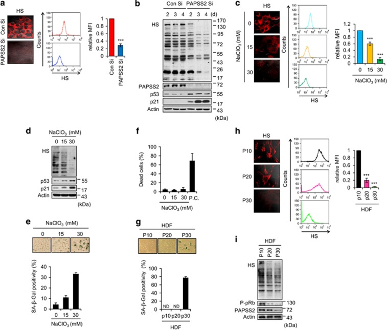
Figure 3.
Decrease in cellular sulfation is associated with premature and replicative senescence. (a–b) MCF7 cells were transfected with either Con Si or PAPSS2 Si. (a) Transfected cells were stained with an anti-HS (10E4) antibody and visualized (left panel) or subjected to FACS to quantify the HS level (middle graph). Quantified data are shown as the mean fluorescence intensity (MFI) relative to Con Si-transfected cells (right graph). (b) Cells were harvested at the indicated times after siRNA transfection and subjected to WB analysis. (c–f) Undersulfation resulting from NaClO3 treatment induces premature senescence. (c) MCF7 cells were cultured in the absence or presence of NaClO3 for 3 d. The HS level were measured as described in a. (d) Analyses of WB, (e) morphological changes and SA-β-Gal positivity, and (f) the percentage of dead cells were performed. (g–i) Cellular sulfation level is associated with replicative senescence in an HDF system. (g) SA-β-Gal positivity in HDFs at passage 10 (p10), p20, and p30. ND, not determined. (h) The HS level was measured as described in a. (i) HDFs were harvested at the indicated passages and subjected to WB analysis. Error bars indicate the S.D. of three independent experiments. ***P<0.001

