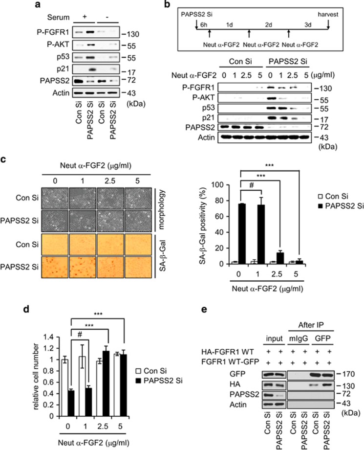
Figure 6.
FGF2 has an essential role in augmented FGFR1-AKT-p53-p21 signaling in PAPSS2-depleted cells. (a) MCF7 cells transfected with Con Si or PAPSS2 Si for 6 h were cultured in the absence or presence of serum for an additional 36 h; cells were then harvested and subjected to WB analysis. (b) MCF7 cells were transfected with Con Si or PAPSS2 Si 6 h before incubation with or without increasing concentrations of Neut α-FGF2 for 3 days (upper timeline). Analyses of (b) WB (lower panel), (c) morphological changes (left panel) and SA-β-Gal positivity (right graph), and (d) relative cell number were performed. Error bars indicate the S.D. of three independent experiments. Statistical significance was determined using one-factor analysis of variance and Holm–Sidak post hoc tests. ***P<0.001 and #P>0.05. (e) Co-IP assay of epitope-tagged FGFR1. Cells were co-transfected with HA- and GFP-tagged WT FGFR1 24 h before transfection with Con Si or PAPSS2 Si. Transfected cells were immunoprecipitated using an anti-GFP antibody and then blotted with an anti-HA antibody

