Figure 2.
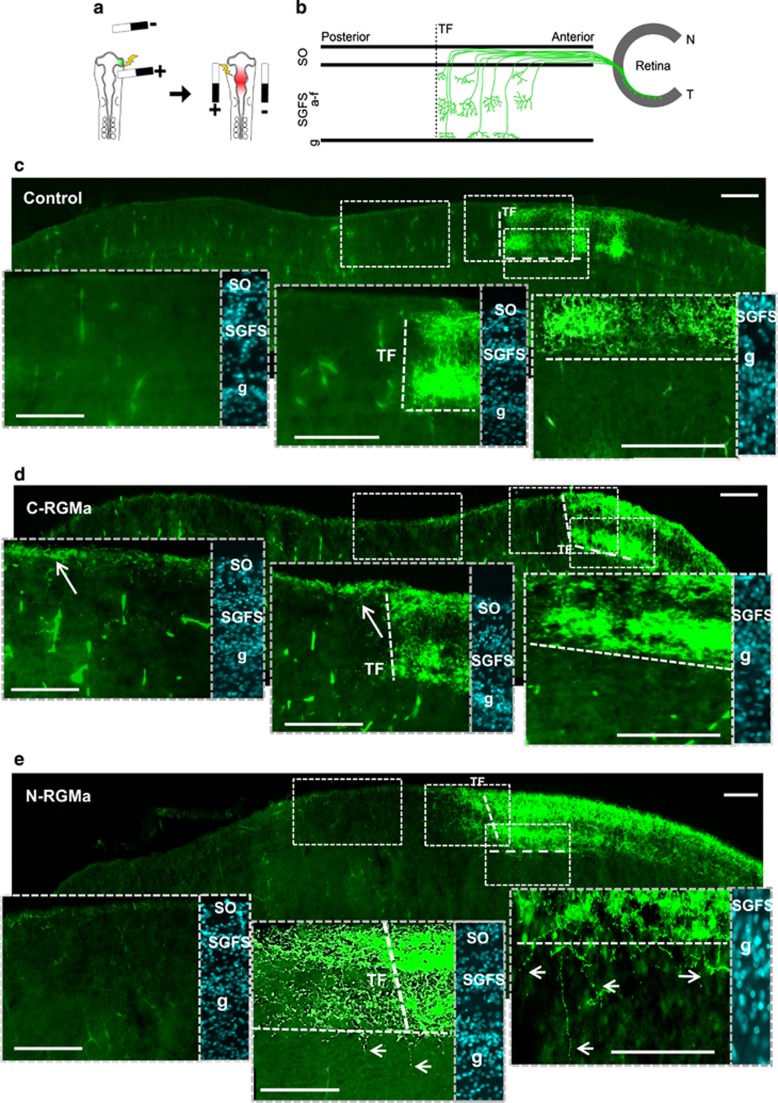
C-RGMa and N-RGMa induce distinct axonal phenotypes. (a) Schematic representation of the experimental approach used to study pathfinding. At first electroporation of the optic vesicle is performed to label temporal fibers with GFP. A second electroporation is performed to allow ectopic expression of a plasmid expressing C-RGMa and N-RGMa in the tectum. (b) Representation of axonal paths from the retina to the SGFS (a–f) laminae of the optic tectum. (c) In controls, temporal axons populate the SO and SGFS layers and terminate precisely at the predicted terminal front (TF; dotted red line). (d) When C-RGMa was overexpressed in the OT, temporal fibers sent overshoots toward the posterior tectum. These overshoots remained restricted to the SO layer (arrow). (e) When N-RGMa was overexpressed in the optic tectum, temporal fibers passed the predicted terminal front (dotted line) and sent overshoots toward the posterior tectum. These overshoots established terminal arbors in the SGFS (a–f) layer. Numerous overshoots crossed the SGFS layer g and were also found in deeper tectal layers (arrows). Bar, 100 μm

