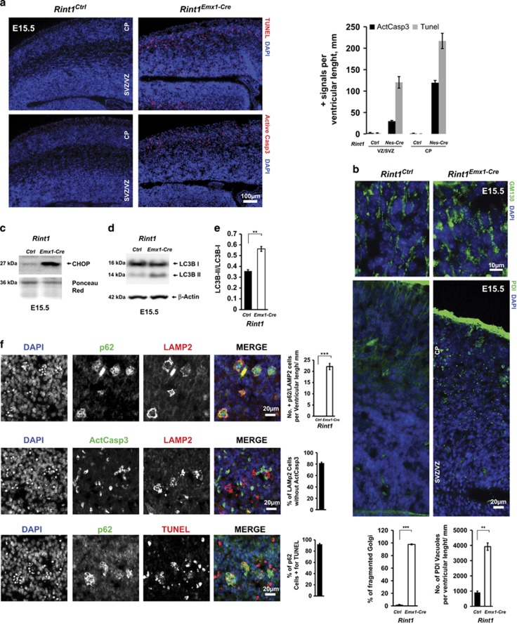
Figure 7.
Deletion of Rint1 in progenitors of dorsal telencephalon cause severe defects already at E15.5. (a) TUNEL and Activated Caspase 3 staining of Rint1Ctrl and Rint1Emx1Cre cortex at 15.5 exhibits extensive apoptosis not only in CP but also in VZ/SVZ as shown in the quantification in the right panel. (b) Destabilization of Golgi (GM130) and ER (PDI) homeostasis in Rint1Emx1Cre cortices with quantification of Golgi fragmentation and ER vacuolization in the bottom panel. Levels of ER stress marker CHOP (c) as well as autophagosome marker LC3B-II (d) are increased in Rint1Emx1Cre cortex. (e) Quantification of LC3B-II/LC3B-I and LC3B-II/Actin ratios in cortex from three different embryos. (f) p62 and LAMP2 accumulation in apoptotic active Caspase3-negative cells in Rint1Emx1Cre cortex at E15.5 with statistic evaluation in the right panel. Stars indicate significance in two tailed Student's t-test **P<0.005, ***P<0.0005

