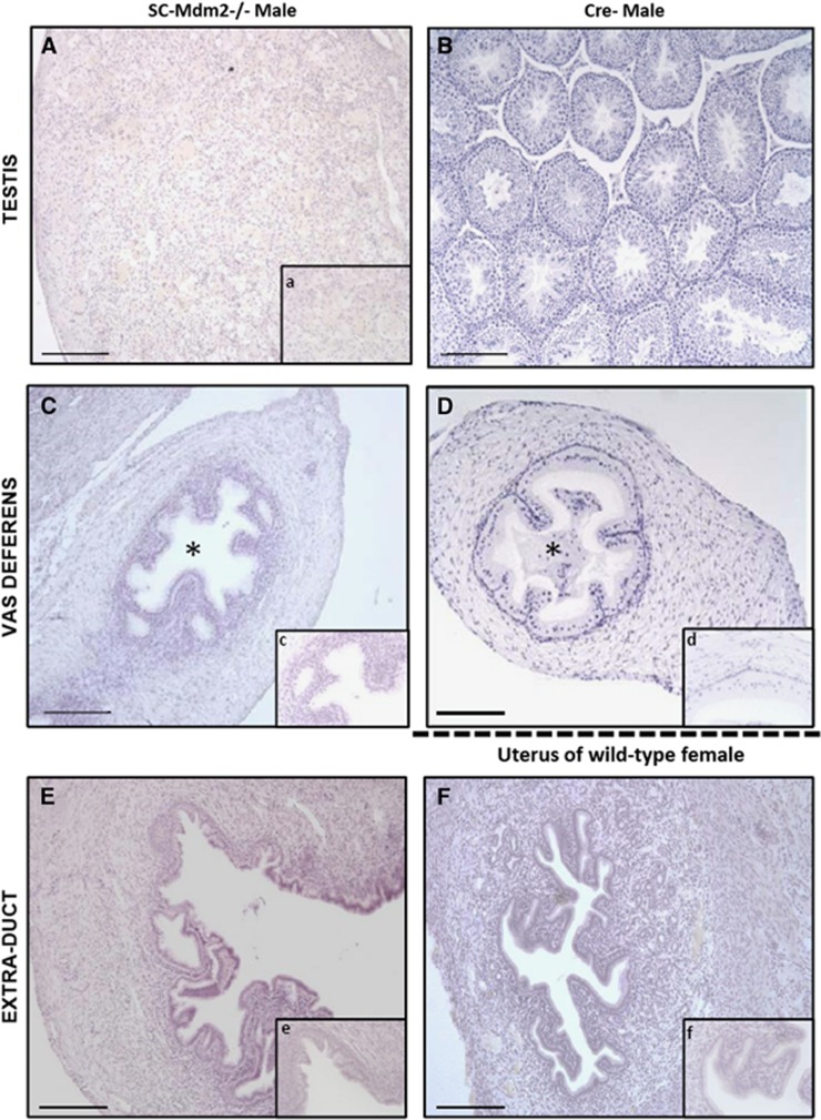
Figure 2.
Histology sections (hematoxylin staining) of testes and ducts in adult SC-Mdm2−/− males (A, C, E) compared with Cre- males for testes and vas deferens (B, C) and females for uterus (E, F). Testicular cells were not organized in seminiferous tubules in adult SC-Mdm2−/− testes (A, a) and no differentiated germ cells were present. No sperm was present in the lumen (*) of the vas deferens contrary to Cre- (C, c and D, d). The histology of the extra duct was similar to the uterus (E, e and F, f). Bar in large windows represents 100 μm; magnification is four times greater in small windows (a, c, d, e, f)

