Figure 4.
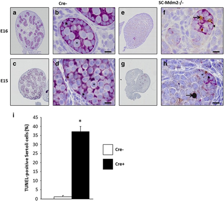
Loss of testicular cords during development is associated with SC apoptosis. Double AMH (purple)/TUNEL (brown) staining was performed with testes at E15 and E16 from control (Cre- a–d) and SCMdm2−/− (e–h) embryos. Left panels (a, c, e, g: low magnification) represent an overview of a representative section of the testes (cartography). Right panels (b, d, f, h: high magnification) represent testicular cords at high magnification. AMH and TUNEL staining were used to reveal apoptotic SCs. A normal cord-like structure was observed in the control testes with SCs surrounding gonocytes. A pronounced loss of AMH staining was observed in SC-Mdm2−/− and the few remaining AMH-positive cells were frequently positive for TUNEL staining as well, indicating that apoptosis occurred mainly in SCs at these stages. Arrowheads point to healthy SCs (b, d and f, h), arrows point to apoptotic SCs (f, h: double purple/brown positive) and asterisks indicate germ cells or gonocytes. Bars represent 10 μm. (i) Measurement of SC apoptosis. The percentage of cells positive for both AMH and TUNEL out of total AMH-positive cells was determined at E15 in Cre- (n=3) and SC-Mdm2−/− (n=5) testis. Data shown as mean +/- S.E.M. Statistical analysis: Mann-Whitney test (*P<0.05)

