Figure 4.

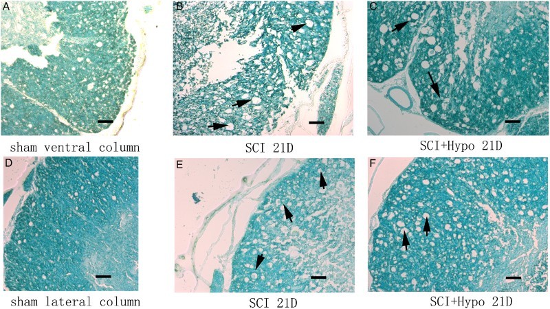
Representative LFB staining of spinal cord (A-C ventral column, D-E: lateral column) on D 21 from the sham, SCI and SCI + hypothermia groups. All black arrows indicate characteristic vacuolation. Scale bar = 200 μm.

Representative LFB staining of spinal cord (A-C ventral column, D-E: lateral column) on D 21 from the sham, SCI and SCI + hypothermia groups. All black arrows indicate characteristic vacuolation. Scale bar = 200 μm.