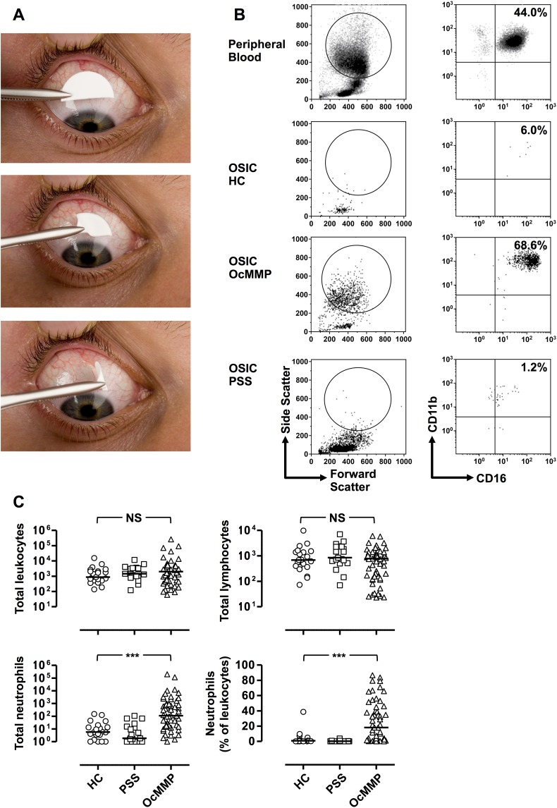
Figure 1.
Ocular mucous membrane pemphigoid is characterized by raised conjunctival epithelial neutrophils. (A) Ocular surface impression cytology was performed with four semicircle membranes per eye (equivalent to two full impression circles) from an anesthetized superior unexposed bulbar conjunctiva. Semicircles were gently pressed in the conjunctiva for a few seconds and lifted with forceps. (B) Representative flow cytometric plots demonstrating the gating strategy to determine conjunctival leukocytes from OSIC and peripheral blood. Live leukocytes were identified by gating for CD45+ cells that were negative for the dead cell exclusion dye Sytox blue. Neutrophils were defined as CD45 intermediate, CD14−, CD16+CD11b+ granulocytes. (C) OSIC-flow of the OcMMP conjunctival epithelium showed predominant neutrophils compared to healthy controls (HC) and primary Sjögren's syndrome (disease controls, pSS). (Comparisons were undertaken by comparing the most inflamed eye at presentation in patients with OcMMP (n = 57) versus the right eye of HC (n = 21) and patients with pSS (n = 19). Three-group comparisons were undertaken by the Kruskal-Wallis test (C) (NS, not significant; *P = 0.01–0.05; **P = 0.001–0.01; ***P < 0.001).

