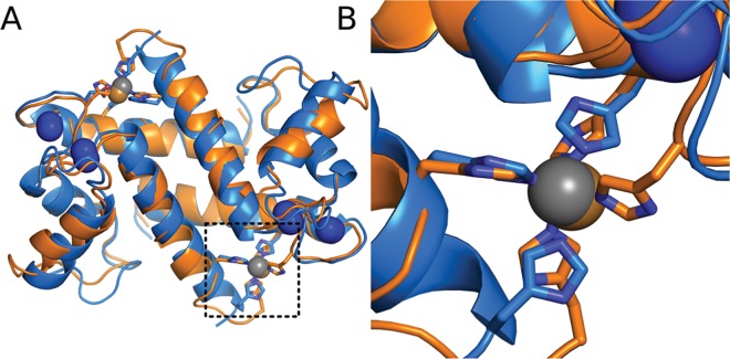
Fig 1. Transition metal binding occurs at a common site in diverse S100 proteins.
Overlay of the crystal structures of S100B (orange, PDB 3CZT) and S100A12 (blue, PDB 1ODB) bound to Ca2+ and transition metals. Ions are shown as colored spheres: Ca2+ (blue), Zn2+ (gray) and Cu2+ (copper). Residues ligating the transition metals are are shown as sticks. Boxed region is shown in detail in panel B.

