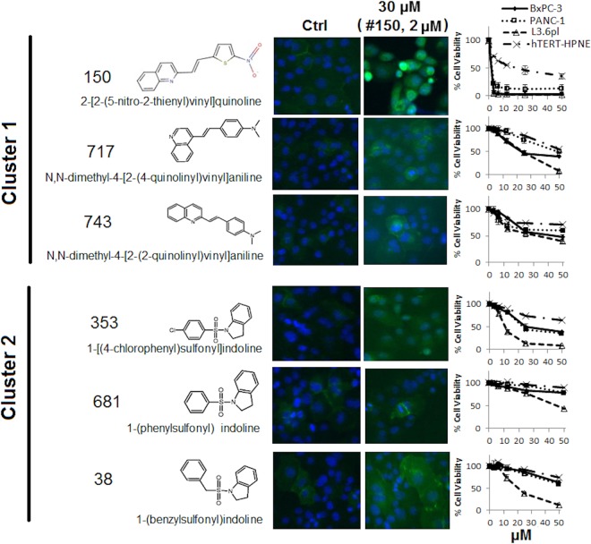
Fig 5. Structure, cytotoxicity and induction of E-cadherin fluorescence by hit compounds.
E-cadherin immunofluorescence of PANC-1 cells treated by 6 selected compounds was detected at 24 hrs of treatment by anti-E-cad primary antibody (1:250 dilution) and Alexa 488 conjugated secondary antibody (1:500 dilution). Sensitivity of PANC-1, BxPC-3, L3.6 and hTERT-HPNE cells to the compounds were detected at 48 hrs treatment by MTT assay. Data represents Mean ± SD of 1–3 independent experiments each done in triplicate.

