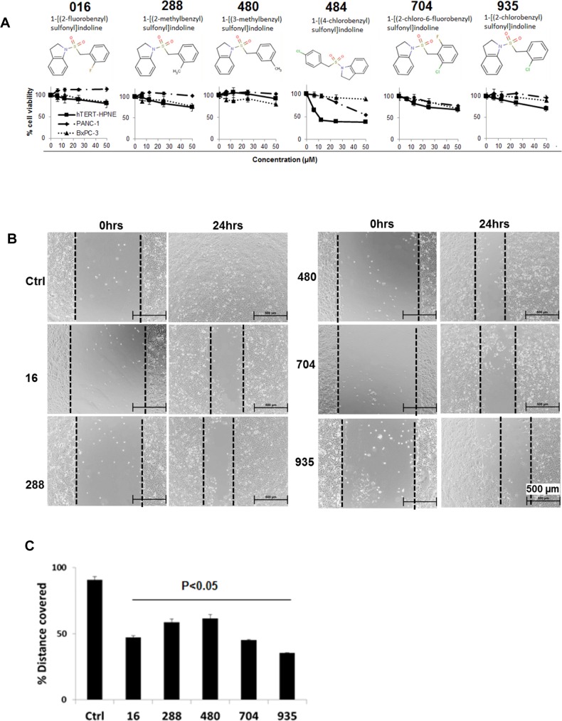
Fig 9. Structure, cytotoxicity and effects of BSI analogues on cell migration.
A. Structure of the 6 BSI analogues, and sensitivity of pancreatic cancer cells (PANC-1 and BxPC-3) cells to BSI analogues. Cells were exposed to different concentrations of BSI analogues for 48 hrs. Cell viability detected at 48hrs post treatment by MTT assay. B. Scratch was made on confluent monolayer of BxPC-3 cells using 1.25 ml sterile pipette tip. After washing with media, cells were exposed to 25 μM BSI analogues. Scratch was photographed at 0 and 24 hrs post treatment. C. Bar graph shows the % distance covered by BxPC-3 cells of 3 repeats.

