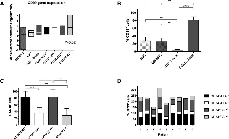
Fig 1. Expression of CD99 in T-ALL and normal hemopoietic cells.
(A) Expression of CD99 antigen was assessed by flow cytometry in BM samples from 9 T-ALL cases and BM MNC, CD34+/CD38- HSC and CD3+ T cells from 5–9 healthy donors. Lines represent median values, ** P≤0.007, **** P<0.0001. (B) Expression of CD99 in sorted LPC subpopulations from this T-ALL cohort. Lines represent median values, * P = 0.02, ** P≤0.009. (C) Stacked bar chart showing expression of CD99 in CD34/CD7 LPC subpopulations in individual cases. (D) Gene expression in BM samples from 5 T-ALL cases (pts 2, 4, 7, 8, 9) and from 5 healthy donors were analyzed using Agilent Whole Genome Oligo microarrays. Data shows side by side comparisons of median centered normalized log2 signal intensities. Boxes represent the range of expression and the horizontal lines represent the median.

