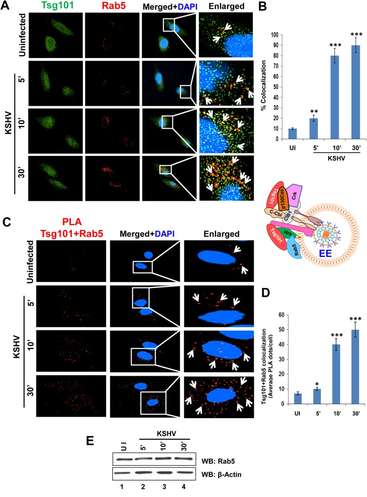
Fig 6. Immunofluorescence and PLA microscopy demonstrating colocalization of TSG101 with early endosome Rab5 during de novo KSHV infection.
Serum starved HMVEC-d cells were infected with KSHV (30 DNA copies/cell) for the indicated time points, washed, and processed for IFA and PLA. Nuclei were stained with DAPI and representative images are shown. Magnification, 40x. Boxed areas from the merged panels are enlarged in the rightmost panels. (A) IFA with mouse anti-Tsg101 (green) and rabbit anti-Rab5 (red) antibodies. White arrows indicate colocalization of Tsg101 with Rab 5 (yellow). (B) Graphical representation of the colocalization shown in 6A as per methods described under the Fig 4B legend. (C) PLA assay using mouse anti-Tsg101 and rabbit anti-Rab5 antibodies. White arrows indicate red spots representing Tsg101 in close proximity with Rab5 positive vesicles. (D) Graphical representation of the average Tsg101-Rab 5 interaction PLA dots per cell shown in 6C. (E) Western blot analysis showing no change in Rab5 protein level upon KSHV infection at the indicated time points.

