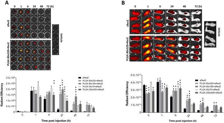
Fig 6. Bioavailability of sRecE and PLGA-RecE in PLNs and injection site.
A) sRecE was tagged with Alexa Fluor 647 and adsorbed to PLGA nanoparticles and inoculated into to footpad. At indicated time points, the draining popliteal lymph nodes (PLNs) were isolated and analyzed for total fluorescence. The upper panel comparing sRecE and 80×320 nm PLGA-RecE is representative for the measure of antigen presence in the PLNs. The comparison of all nanoparticle sizes is depicted in the lower panel. B) The foot pads were analyzed at similar time points to analyze the bio-availability at the inoculation site. Statistical analysis was done by two-way ANOVA followed by Bonferroni posttests. * p<0.05, ** p<0.01, *** p<0.001.

