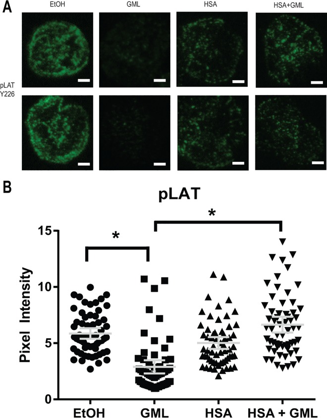
Fig 3. HSA restores GML mediated disruption of LAT microclusters.
(A). APBTs treated with 0.1% ethanol or 10 μg/ml GML in serum free media as well as 0.1% ethanol or 10 μg/ml GML in 1% HSA were stimulated by plate bound anti-CD3 (2 μg/ml) in glass covered chamber slides. They were then fixed, permeabilized, stained with antibody specific for phosphorylated LAT Y226, and imaged using TIRF microscopy. White bar scale indicates 4 μm in length. (B). Pixel intensities of median axis of each cell in images obtained in (A) were quantified and averaged using ImageJ. Scatter plot distributions with 95% confidence intervals of 60 cells from 3 independent experiments are shown. * denotes p<0.05 in Student t’s test comparing the identified samples.

