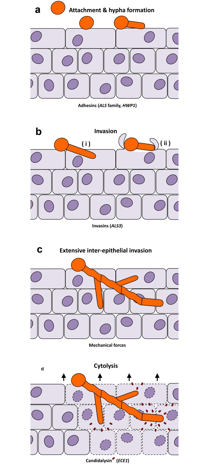
Fig 1. Distinct stages of C. albicans-epithelial infection.
(a) In experimental epithelial infections, C. albicans yeasts form hyphae upon contact with epithelia and adhere tightly to the host cells. This is mediated by a number of adhesins, including members of the Als family and Hwp1. (b) This is followed by initial epithelial invasion via two routes—(i) fungal-driven active penetration and (ii) host-mediated induced endocytosis. (c) Elongating and branching hyphae result in extensive interepithelial invasion. Surprisingly, this invasion itself does not cause damage to the epithelium. (d) Simultaneous secretion of the fungal peptide toxin, Candidalysin (red pentagons), lyses the host epithelia and causes tissue destruction.

