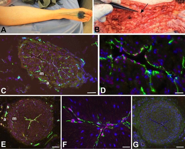
Fig 1. Detection of epifascial lymphatic collectors in patients.
A) Intradermal injection of Patent Blue into the first two interdigital spaces of a patient’s foot. Note the centripetal flow of the marker in the lymphatics. B) Isolation of a lymphatic collector, which is marked blue (arrow), in the hypodermis of the thigh. C-G) Staining with antibodies against CD31 (green) and Prox1 (red) (C-F), and negative control with only secondary antibodies (G). Lymphatic endothelial cell express both of the markers (as seen at higher magnification in D, F). Vasa vasorum in the adventitia (a) and media (m) express only CD31. Bars = 150 μm in C; 25 μm in D; 15 μm in F, and 200 μm in E,G.

