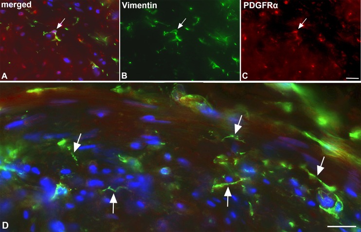
Fig 6. Immunofluorescence studies of ICLC in lymphatic collectors.
Staining with antibodies against vimentin (green) (A, B, D) and PDGFRα (red) (A, C). Note double-positive ramified ICLC (arrow in A-C) in the media of the collector. Bar = 50μm. D) Higher magnification showing vimentin-positive cells with long processes (arrows), which possess varicose-like swellings. Dapi (blue) marks all nuclei. Bar = 35μm.

