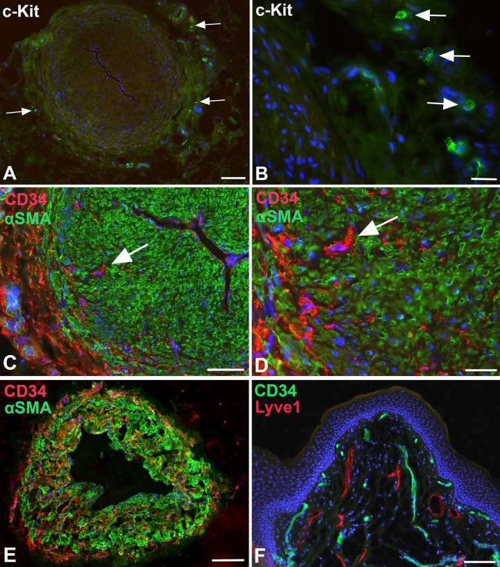
Fig 7. Immunofluorescence studies of lymphatic collectors and dermis.
A, B) Staining with anti-cKIT (CD117) antibodies of lymphatic collectors. Granulated, round cells that express c-KIT (arrows) are located in the adventitia of the collectors. Bar = 250μm in A, and 25μm in B. C-E) Staining of a lymphatic collectors with anti-CD34 (red) and anti-αSMA (green) antibodies. C,D) In large collectors, CD34+ cells (arrow) are found in the adventitia, and in the outer parts of the media between the αSMA-positive SMCs. Bar = 150μm. D) Higher magnification of C showing nucleated CD34+ cells (arrow). No double-positive cells are visible. Bar = 60μm. E) Smaller caliber collector stained with anti-CD34 (red) and anti-αSMA (green) antibodies. CD34+ cells are found in all parts of the media. Bar = 200μm. F) Staining of foreskin with anti-CD34 (green) and anti-Lyve-1 (red) antibodies. Blood capillaries beneath the epidermis express CD34. The Lyve-1-positive lymphatics are CD34-negative. Dapi (blue) marks all nuclei. Bar = 150μm.

