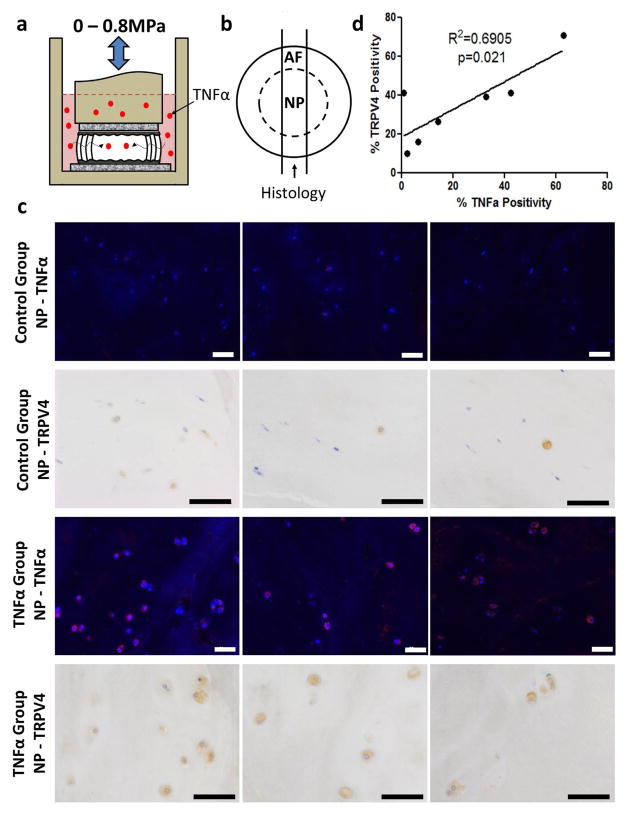
Fig. 2.
TRPV4 is correlated with TNFα expression in an IVD organ culture model of inflammatory degeneration.( a) Schematic of the experimental set-up of the bovine whole IVD organ culture model of degeneration involving TNFα stimulation. (b) Schematic of the transverse cross-section of the IVD demonstrating the location of tissue used for immunohistochemistry and immunofluorescence. c) Immunohistochemical and immunofluorescence staining for TNFα and TRPV4, respectively, in the nucleus pulposus region of the control and TNFα groups. The images in each row are from the same tissue but a different area within each region; scale bar = 50 μm. Staining showed greater TNFα and TRPV4 expression in the TNFα-treated group.( correlation between d)There was a significant corrleation between TNFα and TRPV4 positive cells demonstrating a relationship between TNFα and TRPV4 expression in the IVD. Rabbit control IgG antibody used as a negative control.

