Fig. 6.
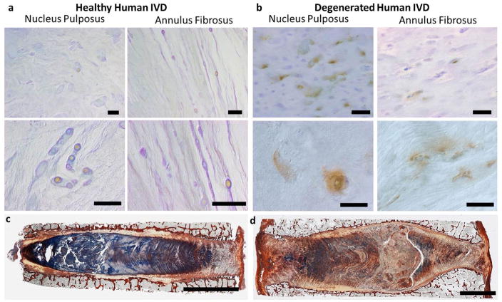
TRPV4 expression in human IVD tissue. Immunohistochemistry for TRPV4 in nucleus pulposus and annulus fibrosus regions of (a) a healthy IVD from a 44 year old male and (b) a degenerated IVD from a 93 year old male; scale bars = 50μm . The images in the top row were taken at low magnification and the bottom row was taken at high magnification under polarised light. Picrosirius red and alcian blue staining of the same (c) healthy 44 year old male IVD and (d) degenerated 93 year old male demonstrating reduced alcian blue staining intensity, indicative of proteoglycan loss, in the degenerated IVD; scale bars = 5 mm. Whole IVD images are modified from (Walter et al., 2014). Results suggest that the changes in TRPV4 expression seen in organ culture and osmolarity experiments are likely to be relevant to the changes that occur in human IVD degeneration.

