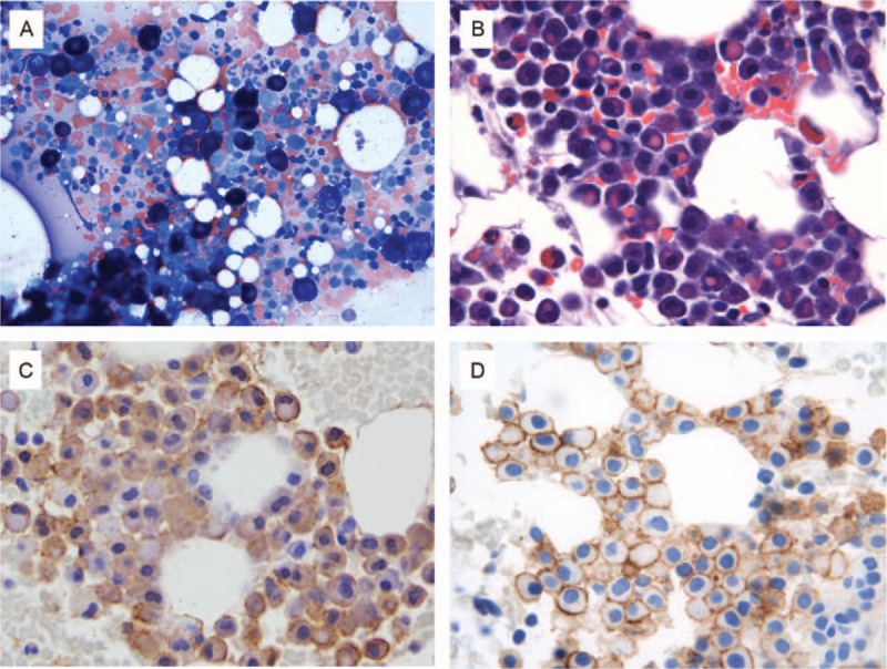
Figure 1.

Histologic and cytomorphologic bone marrow features. (A) Aspirate smear, Wright Giemsa stain 500× magnification; (B) clot section: erythrophagocytosis, hematoxylin, and eosin stain 500× magnification; (C) clot section, mast cell tryptase immunohistochemistry, 500× magnification; (D) clot section, CD30 immunohistochemistry, 500× magnification.
