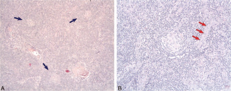
Figure 2.

A, Lymph node histology shows 3 abnormal follicles with hyalinization of germinal center, and an onion-skin aspect of the mantle zone (blue arrows) consistent with hyalin-vascular variant of Castleman disease. B, Higher magnification, centered on the follicle. Blood vessels (red arrows) are visible and are also characteristic of the diagnosis.
