
Figure 2.
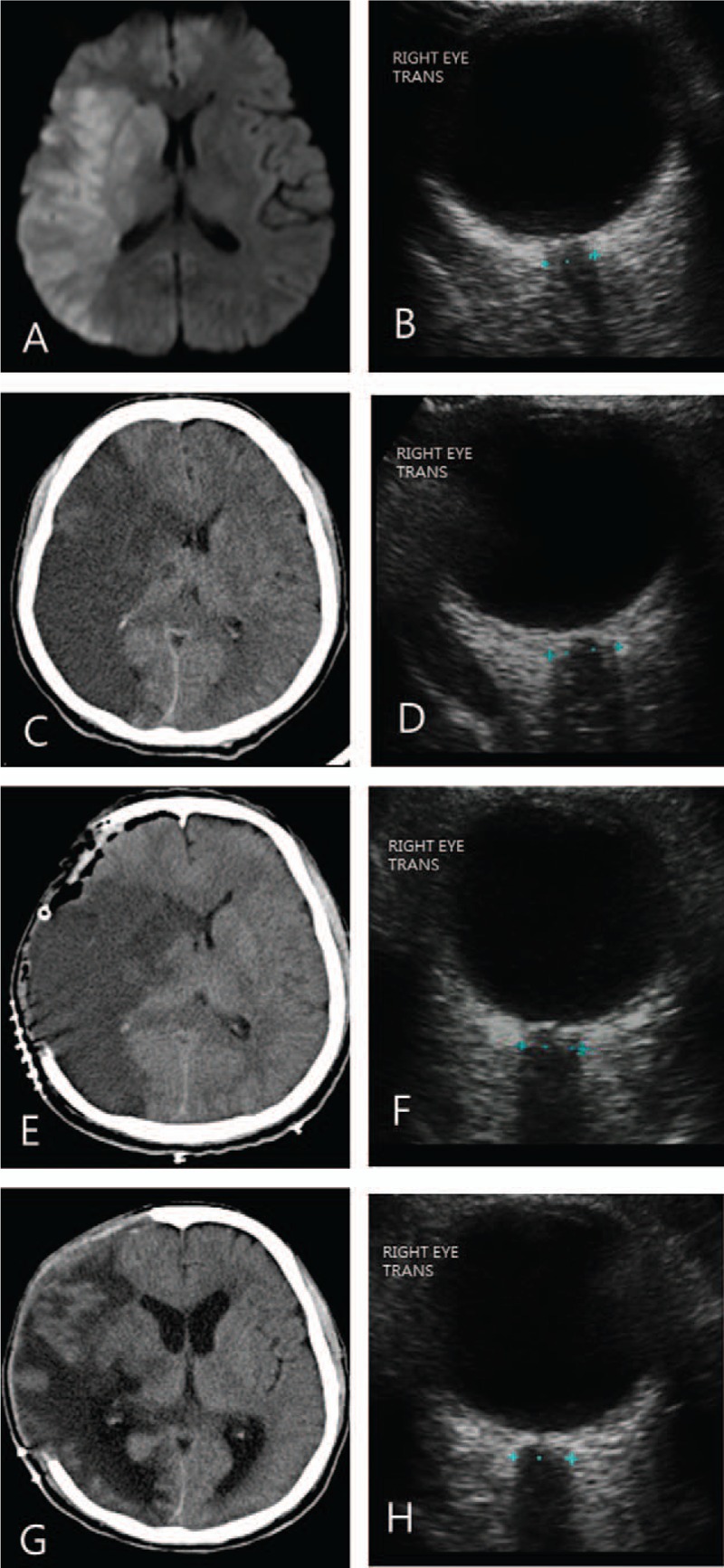
(A, B) A 57-year-old man presented with left hemiparesis because of acute middle cerebral infarction on the right side seen on diffusion magnetic resonance imaging. ONSD on the right side was measured at 5.2 mm. (C, D) The level of consciousness decreased to drowsy on the 2nd day with aggravation of cerebral edema seen on brain CT. ONSD on the right side increased to 6.3 mm. (E, F) Decompressive craniectomy decreased midline shift with ONSD of 5.8 mm. (G, H) CT scans taken after 2weeks after operation showed a substantial improvement in the extent of midline shift with ONSD of 5.4 mm. CT = computed tomography, ONSD = optic nerve sheath diameter.