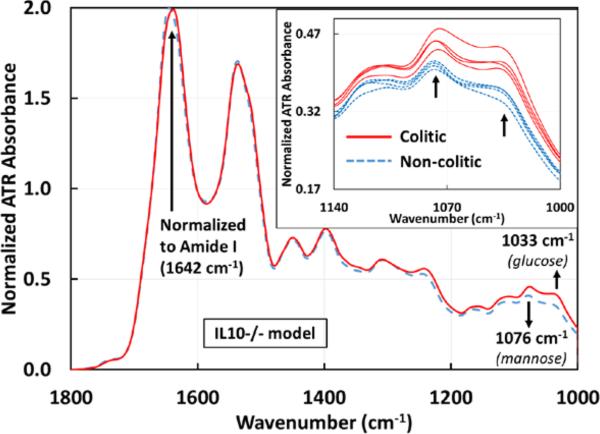
Figure 3.
Averaged ATR-FTIR spectra of sera drawn from IL10−/− mice before (n=4) and after (n=4) spontaneously developing colitis. The same markers 1033 and 1076 cm−1identified in the DSS model are effective in differentiating colitic from non-colitic spectra of the IL10−/− model. The inset shows the individual serum samples from 1140 – 1000 cm−1 for clarity, again showing a clear separation between the two groups. All spectra are normalized to the Amide I peak (1642 cm−1). The averages for the glucose peak are 0.3491±0.0057 (non-colitic) and 0.412±0.009 (colitic) and the averages for the mannose peak are 0.4071±0.0034 (non-colitic) and 0.4553±0.0081 (colitic).

