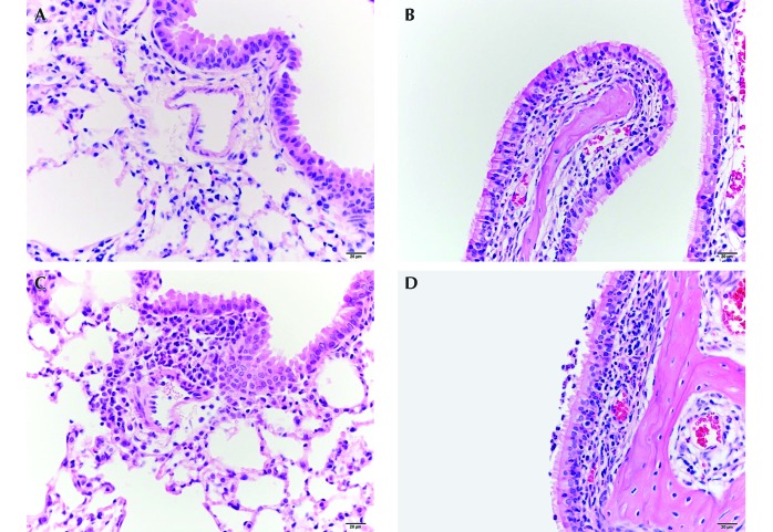
Figure 1.
(A) The lung of mouse 5, which was negative for B. pseudohinzii, shows minimal inflammatory cell infiltrates around pulmonary vessels and bronchioles. (B) The nasal cavity of the same mouse has minimal neutrophilic infiltrates within the respiratory mucosa of the maxilloturbinate. (C) The lung of mouse 2, which was positive for B. pseudohinzii, shows mild inflammatory cell infiltrates (predominantly lymphocytes and plasma cells) around the pulmonary vessels and bronchioles. (D) The nasal cavity of another mouse positive for B. pseudohinzii (mouse 3) has mild neutrophilic infiltration within the respiratory epithelium and lamina propria and minimal neutrophils in the lumen of the lateral meatus.

