Figure 2.
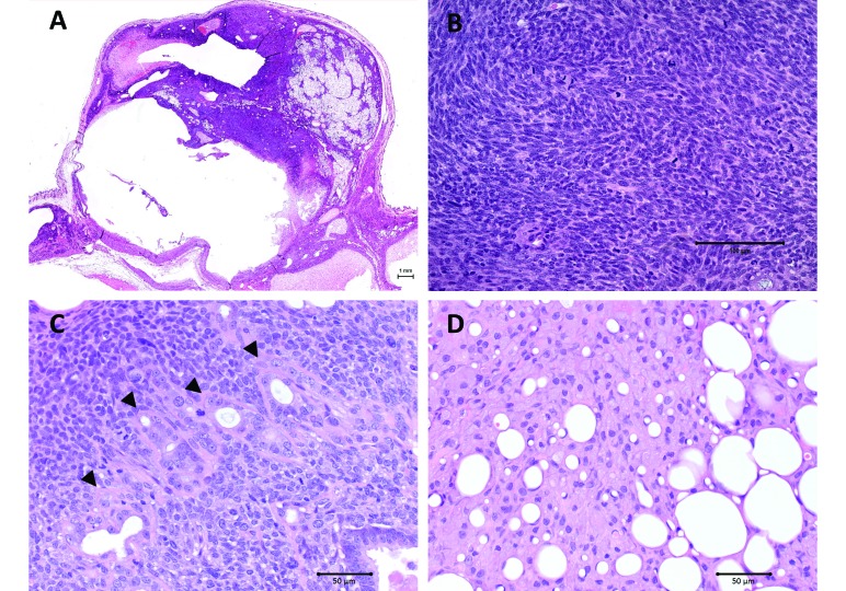
Photomicrographs. (A) Whole-mount cross-section of the mammary tumor. Note seromatous cavity due to prebious FNA (lower left). Bar, 1 mm. (B) Densely cellular neoplastic stroma composed of streams and whorls of mitotically spindle cells with moderate to marked pleomorphism. Magnification, 20×; bar, 100 μm. (C) Malignant epithelial component (arrowheads) comprised of moderately pleomorphic infiltrative-appearing glands. Magnification, 40×; bar, 50 μm. (D) Liposarcomatous element of the neoplasm. Magnification, 40×; bar, 50 μm. Hematoxylin and eosin stain.

