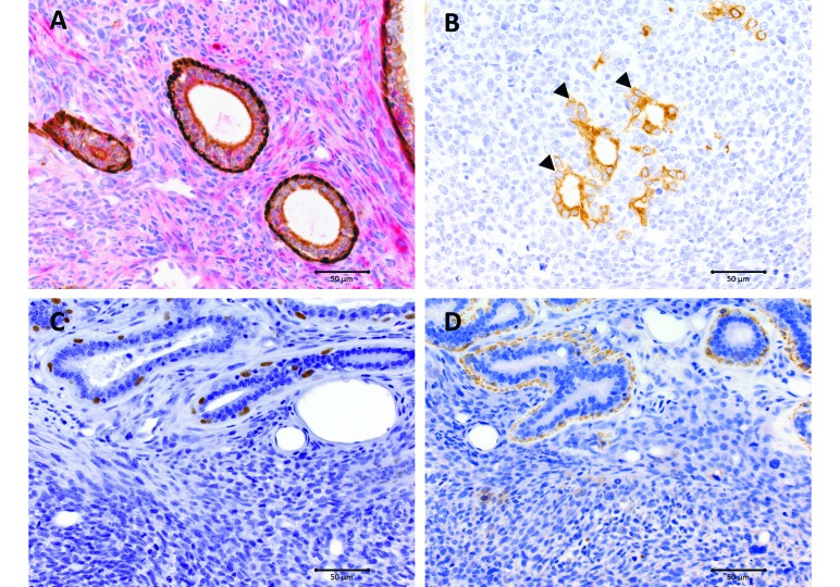
Figure 3.
Photomicrographs of immunohistochemistry. (A) Wide-spectrum cytokeratin and vimentin dual stain. The benign epithelial component is highlighted by cytokeratin staining (brown), with intense nuclear staining of myoepithelial cells and moderate cytoplasmic staining of glandular epithelial cells. The malignant stromal component was vimentin-positive (red). (B) Cytokeratin only. Malignant epithelial component (arrowheads). Note the loss of myoepithelial staining. (C) p63. Nuclear labeling of myoepithelial cells in the benign epithelial component. Neoplastic stromal cells were negative. (D) α-SMA. Cytoplasmic labeling of myoepithelial cells in the benign epithelial component. Neoplastic stromal cells were negative. Magnification, 40×; bar, 50 μm.

