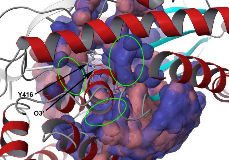
Figure 6.

A comparison between normally solvated structures (red) and structures processed by the water flooding approach (blue). All initial trajectories used were superimposed and the water molecules are presented as molecular surface with transparency. The extra water molecules found around the Y416 after the water flooding are highlighted with circles.
