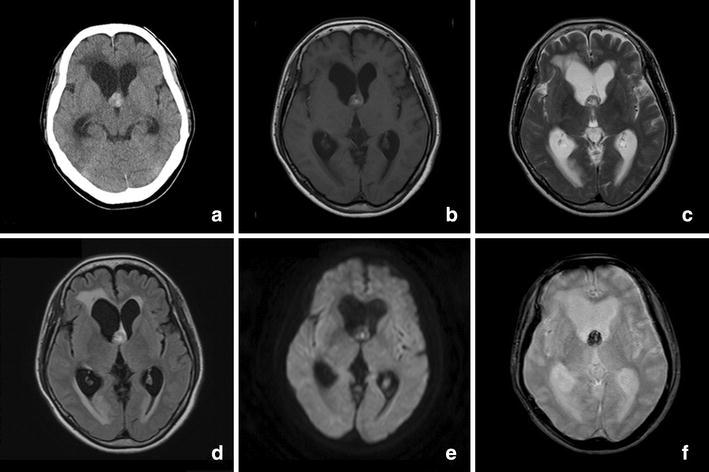
Fig. 1.

Computed tomography and magnetic resonance imagings. a There was a mild hyperintense 16-mm-diameter mass without calcification at the foramen of Monro causing obstructive hydrocephalus. b, c T1- and T2-weighted images showed the well-delineated mixed-signal heterogeneous core. The typical peripheral hemosiderin rim of low signal intensity was not seen on T2-weighted imaging. d No perilesional edema was presented on the fluid-attenuated inversion recovery magnetic resonance image. e Diffusion-weighted imaging showed an isointense mass; only a portion of the mass was hyperintense. f T2-star-weighted imaging showed a hypointense mass
